Autism is a complex disorder of the brain that lasts throughout a
person’s life and affects the ability to communicate, form relationships
with others, and respond appropriately to the environment typically by 3
years of age. In the past two decades autism became a primarily disorder
of brain development. The incidence of Autism Spectrum Disorders is currently
1 in 68 and it affects boys four times more often than girls. As many as
20 or more genes on different chromosomes may be involved in the disorder
to various degrees and determine the severity of symptoms. Interactions between
those genes in addition to environmental factors likely contribute
to variable expression of autism-related traits. The multidisciplinary research
program in my laboratory integrates genetics, molecular and cellular neurobiology
approaches to study the link between the causative biological factors and
behaviour.
Neurobiology of Lipid Signaling in Autism Spectrum Disorders
|
Lipid Components of Cell Membrane Associated
with ASD

The
plasma membrane phospholipids play an important role in the nervous system
and serve as a supply of signaling molecules important for normal functioning
and development of the brain. Research shows that abnormal lipid metabolism
may play a contributing role in the pathology of autism disorders. Lipid homeostasis
can be altered in autism as a result of insufficient dietary supplementation
of omega-6 and omega-3 fatty acids, genetic defects, function
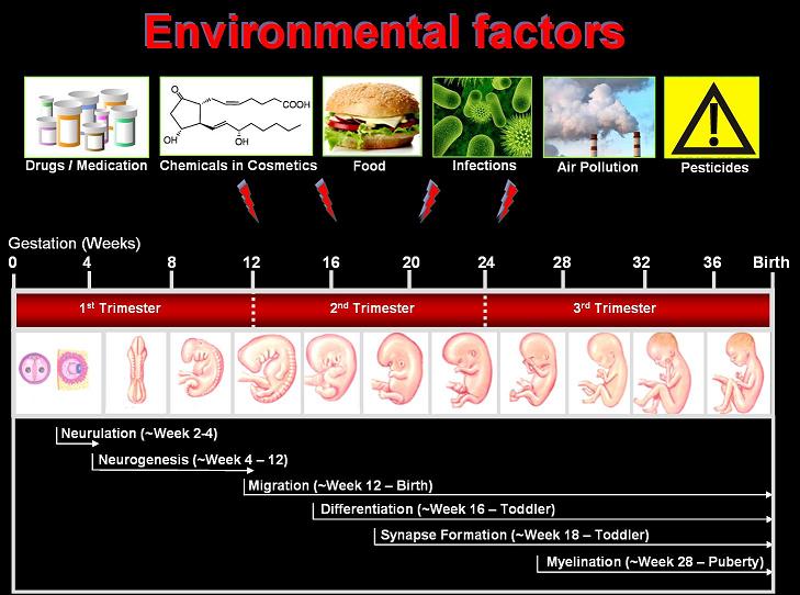
of enzymes involved in their metabolism, or influence of various environmental
agents such as drugs (misoprostol or NSAID), infections or inflammation.
(Dashed arrows indicate an increase or decrease level; asterisks indicate
a link to ASD). (Tamiji
and Crawford 2010a,
Wong et. al. 2013).
|
Defects in Lipid Mediators
Associated with ASD
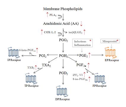
Various bioactive
lipid molecules such as arachidonic acid (AA) can be normally released
from membrane phospholipids by the action of phospholipase A2
(PLA2) and subsequently metabolized into various types of bioactive
prostanoids. Cyclooxygenase-1 enzyme (COX-1), constitutive form, or cyclooxygenase-2
(COX-2), inducible form, converts AA to the unstable PGG2 intermediate
and then to the prostanoid precursor PGH2, which is further metabolized
by the prostaglandin (PG) synthase into the major lipid signaling messengers such as prostaglandins
(PGE2) and other bioactive lipid metabolites
such as prostanoids (PGE2, PGF2α, PGD2,
PGI2) and thromboxane A2 (TXA2).
(Dashed arrows indicate an increase or decrease level; asterisks indicate
a link to ASD). (Tamiji
and Crawford 2010a, Wong et. al. 2013).
|
|
The PGE2 Signalling
Pathway
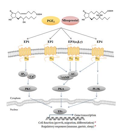
PGE2
diffuses rapidly through membranes, exerting its signalling effects by binding
to E-prostanoid receptors (EP1-4). Evidence shows that it is involved in
early prenatal brain development such as dendritic spine formation, synaptic
plasticity or pain transmission. Clinical studies have revealed a connection
between misuse of the drug misoprostol (an analogue of prostaglandin
type E) during the first trimester of pregnancy and neurodevelopmental aberrations,
including Mobius sequence and ASDs. Misoprostol has been proven to bind and
activate EP receptors activating the PGE2 pathway. During the
early stages of pregnancy (5 to 6 weeks after fertilization), the embryo
is the most vulnerable to misoprostol exposure. We have previously shown that
misoprostol and PGE2 can increase the intracellular level and fluctuation
amplitude of calcium in neuronal growth cones, as well as reduce the number
and length of neurite extensions through the activation of EP receptors.
(Tamiji and Crawford 2010b, Tamiji and Crawford 2010c).
|
Cross-talk between PGE2
and Wnt Signalling Pathways
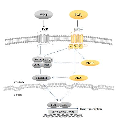
There is growing evidence in non-neuronal cells supporting an interaction between the PGE2 and the Wnt (wingless) pathways. Such an interaction is of particular interest since Wnts are morphogens necessary for the formation of a healthy nervous system. Our lab investigates the interaction between these two pathways in the nervous system. We have shown that PGE2 can modulate the expression of Wnt-target genes (Ctnnb1, Ptgs2, Ccnd1, Mmp9) and change the Wnt-dependent proliferation and migration behaviour of neuroectodermal (NE-4C) stem cells. We also show that PGE2 treatment leads to earlier formation of neural stem cell clusters called neurospheres during differentiation. PGE2 also changed the expression of Wnt pathway genes during cell differentiation, including Wnt2, Wnt3, Wnt8a, Tcf4, Ccnd1, Mmp2, and Mmp9. All these genes have been linked to neurodevelopmental disorders, including ASD. (Wong et. al. 2014) |