An internet resource developed by
Christopher D. Green
York University, Toronto, Ontario
ISSN 1492-3173
Need [AS. nyd]: Ger. Bedürfniss; Fr. besoin; Ital. bisogno. A constitutional or acquired craving or want, either bodily, revealing itself also in consciousness, or mental.
Needs are deep-seated demands of nature; appeased by recurrent satisfactions;
extremely painful or depressing if not satisfied; and often acting as subconscious
motives which influence action without taking form as conscious ends. (J.M.B.,
G.F.S.)
Negation [Lat. negatio, which translates Gr. apofasiV]: Ger. Verneinung; Fr. négation; Ital. negazione. Negation is used (1) logically, (2) metaphysically. In the logical sense it may be used (a) relatively, and (b) absolutely. Used relatively, when applied to a proposition, it may be understood (a) as denying the proposition, or (b) as denying the predicate.
(1) In its logical sense, negation is opposed to affirmation, although, when it is used relatively, this is perhaps not a convenient contrary term; in its metaphysical sense, negative is opposed to positive (fact, &c.).
The conception of negation, objectively considered, is one of the most important of logical relations; but subjectively considered, it is not a term of logic at all, but is pre-logical. That is to say, it is one of those ideas which must have been fully developed and mastered before the idea of investigating the legitimacy of reasonings could have been carried to any extent.
The treatment of the doctrine of negation affords a good illustration of the effects of applying the principle of PRAGMATISM (q.v.) in logic. The pragmatist has in view a definite purpose in investigating logical questions. He wishes to ascertain the general conditions of truth. Now, without of course undertaking to present here the whole development of thought, let it be said that it is found that the first step must be to define how two propositions can be so related that under all circumstances whatsoever,
This must be the first part of logic. It is deductive logic, or (to name it by its principal result) syllogistic. At all times this part of logic has been recognized as a necessary preliminary to further investigation. Deductive and inductive or methodological logic have always been distinguished; and the former has generally been called by that name.The truth of the one entails the truth of the other,
The truth of the one entails the falsity of the other,
The falsity of the one entails the truth of the other,
The falsity of the one entails the falsity of the other.
In order to trace these relations between propositions, it is necessary to dissect the propositions to a certain extent. There are different ways in which propositions can be dissected. Some of them conduce in no measure to the solution of the present problem, and will be eschewed by the pragmatist at this stage of the investigation. Such, for example, is that which makes the copula a distinct part of the proposition. It may be that there are different ways of useful dissection; but the common one, which alone has been sufficiently studied, may be described as follows:
Taking any proposition whatever, as 'Every priest marries some woman to some man,'
we notice that certain parts may be struck out so as to leave a blank form, in which, if the blanks are filled by proper names (of individual objects known to exist), there will be a complete proposition (however silly and false). Such blank forms are, for example:
Every priest marries some woman to ______,
_____ marries _____ to some man,
_____ marries _____ to _____.
It may be that there is some language in which the blanks in such forms cannot be filled with proper names so as to make perfect propositions, because the syntax may be different for sentences involving proper names. But it does not matter what the rules of grammar may be.
The last of the above blank forms is distinguished by containing no selective word such as some, every, any, or any expression equivalent in force to such a word. It may be called a PREDICATE (q.v., sense 2) or rhma. Corresponding to every such predicate there is another, such that if all the blanks in the two be filled with the same set of proper names (of individuals known to exist), one of the two resulting propositions will be true, which the other is false; as
Chrysostom marries Helena to Constantine;
Chrysostom non-marries Helena to Constantine.
It is true that the latter is not good grammar; but that is not of the smallest consequence. Two such propositions are said to be contradictories, and two such predicates to be negatives of one another, or each to result from the negation of the other. Two propositions involving selective expressions may be contradictories; but in order to be so, each selective has to be changed from indicating a suitable selection to indicating any selection that may be made, or vice versa. Thus the two following propositions are contradictories:
Every priest marries some woman to every man;
Some priest non-marries every woman to some man.
It is very convenient to express the negative of a predicate by simply attaching a non to it. If we adopt that plan, non-non-marries must be considered as equivalent to marries. It so happens that both in Latin and in English this convention agrees with the usage of the language. There is probably but a small minority of languages of the globe in which this very artificial rule prevails. Of two contradictory propositions each is said to result from the negation of the other.
The relation of negation may be regarded as defined by the principles of contradiction and excluded middle. See LAWS OF THOUGHT. That is an admissible, but not a necessary, point of view. Out of the conceptions of non-relative deductive logic, such as consequence, coexistence or composition, aggregation, incompossibility, negation, &c., it is only necessary to select two, and almost any two at that, to have the material needed for defining the others. What ones are to be selected is a question the decision of which transcends the function of this branch of logic. Hence the indisputable merit of Mrs. Frankin's eight copula-signs, which are exhibited as of co-ordinate formal rank. But, so regarded, they are not properly copulas or assertions of the relation between the several individual subjects and the predicate, but mere signs of the logical relations between different components of the predicate. The logical doctrine connected with those signs is of considerable importance to the theory of pragmatism.
For the negation of modals see MODAL.
Conversion by negation = CONTRAPOSITION (q.v.).
Negant or negative negation is the negation effected by attaching the negative particle to the copula in the usual Latin idiom, 'Socrates non est stultus,' in contradistinction to infinite (aoristh), or infinitant, negation, which is effected by attaching the negative particle to the predicate, 'Socrates est non stultus.'
Kant revived this distinction in order to get a triad to make out the symmetry of his table of categories; and it has ever since been one of the deepest and dearest studies of German logicians. No idea is more essentially dualistic, and distinctly not triadic, than negation. Not-A = other than A = a second thing to A. Language preserves many traces of this. Dubius is between two alternatives, yea and nay.
(2) In the metaphysical sense, negation is the mere absence of a character or relation that is regarded as positive. It is distinguished from privation in not implying anything further.
Spinoza's celebrated saying, of which the Schellings have made so much, 'omnis determinatio est negatio,' has at least this foundation, that determinatio to one alternative excludes us from another. The same great truth is impressed upon youth in the utterance: 'You cannot eat your cake and have it too'. (C.S.P., C.L.F.)
Predicates are not denied to subjects at hazard -- it would be a great waste
of time to set forth in language the fact that the vast majority of predicates
are inapplicable to the vast majority of subjects. In order that a negative
statement may have any value, there must have been some reason to suppose that
the affirmative statement of which it is the exact denial was true, either that
it had been proposed for our acceptance by an interlocutor, that it had been
part of our stored-up knowledge or purported knowledge, or that we had in mind
what we took at the moment to be sufficient ground for its acceptance. Sigwart
is, therefore, right in maintaining that the negative statement, in its origin,
is not of the same primitiveness as the affirmative statement; 'a is
not b' is merely a shorter form, permitted by language, for 'that a
is b is false,' or 'that a is b is non-occurrent.' Cf.
JUDGMENT, ad fin. (C.L.F., J.M.B.)
Negative [Lat. negativa; a term appearing first in logic in Boethius, in place of the previous abdicativa, although negatio was much earlier. It translates Aristotle's apofatikh. Cognate words were used by Plato, and even earlier]: Ger. verneinend; Fr. négatif; Ital. negativo. Involving NEGATION (q.v.), either in the second application of the logical sense, or in the metaphysical sense given under that term.
Negative abstraction is an act of abstraction derived from considering something which does not possess the character considered.
Negative (or necessary) condition: see NECESSARY AND SUFFICIENT CONDITION.
Negative criterion: a criterion which is a negative condition; a test. Most criteria are of this sort.
Negative discrepancy: see DISCREPANCY.
Negative distinction: a mutual real distinction separating anything from its negation; as the distinctions of heat and cold (no heat), light and darkness (no light), sound and silence (no sound).
Negative idea: see Negative name.
Negative mark: a mark which consists in the non-occurrence of a positive phenomenon under certain conditions.
Negative name: a common name which characterizes an object by its want of some character. 'I appeal,' says Locke, 'to every one's own experience, whether the shadow of a man, though it consists of nothing but the absence of light (and the more the absence of light is, the more discernible is the shadow), does not, when a man looks on it, cause as clear and positive an idea in his mind as a man himself, though covered over with clear sunshine? And the picture of a shadow is a positive thing. Indeed, we have negative names, which stand not directly for positive ideas, but for their absence, such as insipid, silence, nihil, &c., which words denote the positive ideas, taste, sound, being, with a signification of their absence' (Essay concerning Human Understanding, II. viii. 5).
Negative negation: see NEGATION.
Negative syllogism: any syllogism of the second figure, or the modus tollens, where the reasoning turns upon the change of quality. The canon of syllogism, that nothing can be concluded from two negatives, is inaccurate. What is requisite, in non-relative syllogism, is that the middle term should be once distributed and once undistributed. Darapti and Felapton, which appear to violate this rule, only do so because one of the premises, so far as it is efficient, is virtually a particular. What is requisite is, that one of the interlocutors should select the individual denoted by the middle term in one premise and the other in the other.
Negative whole is one which has no parts; as God, the soul, &c. (C.S.P.)
Negative term. Negation arises first, without doubt, in connection with
the judgment -- it is a secondary function of thought, which presupposes the
existence of positive judgments (Hamilton, Sigwart, Wundt). It is true that
the concept cat cannot be formed by the child except by separating out a certain
quality-complex from a background of all that is other than cat; but this background
exists in its mind only vaguely, like images upon a retinal periphery, and until
it has become a distinct object of consciousness it does not constitute a concept.
Later, thought permits itself to affirm not only that a is-not b, but
also that a is not-b. The concept not-b is, in many cases, no
more difficult to form than the concept b; it is frequently hard to say
which of two concepts, as odd or even, to greet an acquaintance or to cut him,
is positive and which is negative -- to be immortal means to continue to live,
and to be mortal means not to continue to live. But this similarity between
positive and negative terms holds only so long as the quality which constitutes
their signification is one and indivisible. Terms in general are implements
for holding together a certain group of objects, each in the possession of a
certain complex of marks; a negative term has for its denotation all other
objects in the universe of discourse, whatever that may be, and for its connotation
the absence of some one at least of the elements of the complex of marks
signified by the positive term. The group of objects to which a negative term
applies is all the objects other than those to which the positive term applies;
for signification there is not, it is true, any mark common to this group of
objects (for in general they have no such mark), but this is merely to say that
a negative name has no positive concept corresponding to it (Keynes). The significations
of the positive term and of the negative term are very different; the one involves
a combination of quality-elements, the other an alternation of absences
of quality-elements. When, therefore, Lotze says that it remains a forever insoluble
task to abstract the qualities of the not-man, he says what is true but
unimportant. Not-man is not destitute of import, as Lotze says it is,
but its import consists in an alternation of deficiencies of some one,
at least, of the elements of the intent of man. (C.L.F.)
Negative Sensation: Ger. negative Empfindung; Fr. sensation négative; Ital. sensazione negativa. Sensation due to stimulus beneath the THRESHOLD (q.v.).
Used by Wundt (Physiol. Psychol., 3rd ed., 384), who gives the
negative sensation a greater value as the stimulus continues to sink, until
at zero stimulus the negative sensation is infinite, and is 'more undetectable
than any other undetectable sensation.' All of which, in the opinion of some,
in reine Mythologie. (J.M.B.- G.F.S.)
Negative Variation: Ger. negative Schwankung, Actionsstrom; Fr. variation négative; Ital. variazione negativa. Decrease of the muscle or nerve current upon tetanizing an excised muscle or nerve.
Since any portion of a muscle or nerve which is active becomes electrically negative to resting portions, the term 'action current' is to be preferred to negative variation, and has come into general use in its stead.
The phenomenon was discovered independently by Matteucci and Du Bois-Reymond,
1840-3. (C.F.H.)
Negativism: see UNKNOWABLE, and cf.
AGNOSTICISM. See also PSYCHOSIS.
Negativity and Negation (Hegel's
Negativität and Negation): see HEGEL'S TERMINOLOGY,
Glossary, sub verbis.
Negligence (in law) [Lat. negligentia]: Ger. Fahrlässigkeit, Nachlässigkeit; Fr. faute, négligence; Ital. negligenza. Acting or omitting to act without due care to avoid or prevent injury to others.
Due care is that which, under the circumstances of the particular case, is required by the rules of law. It may, by one of these rules, be such care as a man of ordinary prudence in such a situation would reasonably be expected to use. To constitute actionable negligence there must be an obligation towards the party who claims the right of action to use care, and a breach of that obligation to his injury (Markby's Elements of Law, § 681; Holland, Jurisprudence, chap. viii. 98, 99). Contributory negligence is a bar to an action for negligence, and means negligence of the plaintiff materially contributing to produce the injury of which he complains. Gross negligence is the absence of even slight care. 'Gross negligence is not actionable where not even slight care was due. However blameworthy, it is still essentially different from intentional wrongdoing' (Beers v. Boston and Albany Railroad Co., 67 Conn. Law Reports, 427). Under the doctrine of Respondeat superior, a master may be liable for his servant's negligence, and may be liable for negligence in employing a careless or incompetent servant, by whose act another suffers.
Negligentia does not seem to have been actionable, at Roman law, unless
it so infringed upon the rights of others as to constitute culpa. 'Magna
negligentia culpa est (Dig., 1. 16, de verborum significatione,
226). See Cushing, Introd. to Roman Law, § 369 ff.; Morey,
Outlines of Roman Law, 349. (S.E.B.)
Nemesis [Gr. nemesiV, from nemein, to distribute or allot]: Ger. Nemesis; Fr. Némésis; Ital. nemesi. In Greek thought, the principle of inevitable retribution according to the strict measure of desert; conceived at first impersonally, but later personified as a divinity.
Nemesis is distinguished from Fate, which in Greek thought has scarcely any
moral significance. Nemesis is the vengeance which man brings upon himself by
his own deeds. In this sense it corresponds to Karma in Hindu thought. There
is no escape from Nemesis, however, whereas Hinduism points out a way of salvation.
(A.T.O.)
Nemesius: see PATRISTIC PHILOSOPHY, (6)
(c).
Neo- [Gr. neoV, new]:
Ger. Neo-; Fr. néo-; Ital. neo-. New; as in neophobia,
an abhorrence of anything new; neomania, a passion for novelty. (J.J.)
Neo-criticism: Ger. Neo-Kriticismus;
Fr. néocriticisme; Ital. neo-criticismo. The revived KANTIANISM
(q.v.) of the 19th century; theoretically equivalent to neo-Kantianism, but
as matter of fact used mainly for the form given to the Kantian thought in France
by Renouvier, Pillon, and others. (J.D.)
Neo-Hegelianism: Ger. Neuhegelismus; Fr. néo-hégélianisme; Ital. neo-Hegelianismo. The revived Hegelianism of the school of thinkers represented in England by T. H. Green, J. and E. Caird, and Bosanquet, and in the United States by Morris, Harris, and Dewey.
The spirit of Hegel and the general outcome of his idealism are revived rather
than the actual developments of his thought or his dialectical method. (J.M.B.)
Neo-Kantianism: Ger. Neo-Kantianismus;
Fr. Néo-Kantisme; Ital. Neo-Kantianismo. See KANTIANISM,
and NEO-CRITICISM.
Neo-Lamarckism: see LAMARCKISM.
Neology [Gr. neoV,
new, + logoV, science]. Used in theology in a sense
identical with RATIONALISM (q.v.). (A.T.O.)
Neo-Platonism: Ger. Neuplatonismus; Fr. néoplatonisme; Ital. neo-platonismo. (1) The revival and transformation of Platonic philosophy that took place, with Alexandria as its head quarters, under the influence of oriental thought. Cf. Whittaker, The Neo-Platonists (1901). See PATRISTIC PHILOSOPHY (2), ALEXANDRIAN SCHOOL, and SOCRATICS (Plato).
(2) Also, the revival of Platonism that took place at Cambridge, England, in
the 17th century, under the influence of Cudworth and Henry More. See CAMBRIDGE
PLATONISTS. (J.D.)
Neo-Pythagoreanism: Ger. neupythagoreische Lehre (K.G.); Fr. néo-Pitagoreisme; Ital. neo-pitagoreismo. A system or, better, a tendency of thought which arose at Alexandria in the 1st century A.D., and which, in accordance with the tendency of the time, tried to consecrate its own teachings by identifying them with the teaching of some ancient sage.
It had its derivation from the latest period of the philosophy of Plato, in
which the categories of the One, the Dyad, the Odd and Even, &c., were manipulated;
and it emphasized the dualism of the Platonic metaphysic, transforming it into
the basis of an ethical and religious asceticism. In connection with the latter
it revived also the Pythagorean mysteries. It is of chief philosophic import
because of its influence, upon Clement and Origen. Cf. PATRISTIC PHILOSOPHY.
(J.D.)
Nerve [Lat. nervus]: see NERVOUS SYSTEM
(III), and NERVE STIMULATION AND CONDUCTION.
Nerve-cell: see NERVOUS SYSTEM (II).
Cf. NEUROCYTE, and NEURONE.
Nerve Stimulation and Conduction: (foreign equivalents are given under STIMULATION, and CONDUCTION). All nervous structures, whether fibres or cells, have normally the twofold property of reacting upon stimuli of suitable kinds (stimulation) and of propagating the peculiar form of molecular activity thus set up to adjacent or connected nerve elements (conduction).
It might be supposed that the cells would prove to be pre-eminently the excitable elements and the fibres the conducting members, but no such complete differentiation has taken place. Neither is it possible completely to distinguish these two processes one from the other, for the process of excitation is not different in kind from that which is propagated from one point to another in the system.
It is one of the commonest facts of nerve physiology that there is some peculiarity developed in the mature nerve-fibre which strongly tends to polarize its elements in such a way as to make more easy the passage of a stimulus in one direction than in the opposite one. It is not to be doubted that this peculiar resistance to translation of the stimulus or current the wrong way depends on some peculiarity of what is called molecular arrangement, and it is also more than probable that this structural organization is a function of the earliest activities of the axis cylinder which transform the before indifferent protoplasm into the forward-conducting nerve. The curious relation of this directive polarity of nerves to their original direction of growth is noted under NERVOUS SYSTEM (q.v., III). See also REGENERATION. The various experimental attempts to prove the power of nerves to conduct in the reverse direction suffer from the fact that all evidence shows that the union of an afferent and efferent nerve cannot take place without a regeneration of the axis cylinders beyond the lesion or point of attempted union.
In general it may be said that under favourable circumstances nearly all kinds of molar and molecular stimuli are capable of producing an effect on nervous matter. See PROTOPLASM, and LIVING MATTER.
One of the simplest means of experimental examination of the conductivity of nerves is that afforded by the so-called nerve-muscle apparatus, too well known to require description here. By varying the experiments with this apparatus it is possible to determine approximately the time required for a nerve-current to pass through a definite length of the nerve and numerous other data respecting the conductivity of the nerve. These experiments are all, of course, complicated by the fact that the current must produce a muscular contraction before a registration can be secured, and numerous variables may exist while the observer is attempting to measure one. By the use of various forms of electrometer, nervous responses may be studied in terms of the electrical variation accompanying them. Here, too, the greatest caution is required in interpreting the results. While shock, thermal irritation, and chemical stimuli all produce nervous activity, the application of the various forms of electrical stimuli is so much more accurate and practicable that nearly all work in the study of nervous conduction is done by such means.
Du Bois-Reymond, who is the pioneer in the study of these phenomena, formulated in 1845 the statement that the excitory effect of a constant current is secured only by varying the intensity of the current; an unvarying constant current does not act as an exciter. The resistance offered by living nerve to the current is about the same as that of muscle. It is, in round numbers, 50,000,000 as great as that of copper wire, and 14.86 times that of distilled water. A current impinging on a nerve-fibre at right angles to the course of the nerve produces no effect, and the effect varies with the direction (ascending or descending), with the duration of the stimulus, the strength of the stimulus, and the length of the nerve through which it flows. The minimum time during which the current must be applied is apparently about .0015 of a second. When one electrode is placed at the equator and the other at the cut end of a nerve, the needle of a sensitive galvanometer indicates the passage of a so-called current of rest. But now, if an interrupted current be applied to the nerve, awakening its physiological activity, the needle of the galvanometer moves backward towards zero. This 'negative variation' may be produced by any stimulus that will set up a nervous action in the fibre. The electro-motive force of a nerve is thus lessened by its physiological activity, and the amount of diminution finds its measure in the negative to all the places in the same nerve-fibre that are not excited. Du Bois-Reymond stated that the negative variation is not a continuous lessening, but a successive rise and fall of the current.
In his early experiments (1871) Bernstein made the following points: Between the moment of stimulation of a given point in a nerve and the onset of negative variation at a given distance in that nerve a measurable time elapses. The phenomena of negative variation have also a measurable duration. The curve of negative variation rises abruptly to its maximum, but sinks more gradually. The rate of translation of the stimulus and that of the negative variation are identical. The duration of the negative variation was found to lie between .0006 and .0007 sec., the length of the wave being 18 mm., and the rate of translation 28 metres per second. With strong stimuli the negative variation may greatly exceed the nerve-current. The duration of a wave 10 mm. long in muscle is .004 sec., and the rate 3 to 4 metres. In nerves the wave remains more or less constant in its passage from the point of excitation, but in muscles, because of the transformation of molecular into molar motion, the intensity of negative variation diminishes.
In 1836 Peltier described a negative polarization in the legs of a frog through which a current was passing. Du Bois-Reymond found the point where the current entered alkaline, and that where it emerged acid. It then appeared that any porous body not too poor a conductor would, under similar circumstances, exhibit this negative polarization. Matteucci in 1860 independently described the phenomena of negative polarization and termed them secondary electromotor phenomena. In addition to the negative polarization, which at first does not seem to differ from that of other porous conducting bodies, but really is somehow connected with the vital activities of the tissue, there is a positive polarization which, so far as known, is confined to nerve, muscle, and electric organs. In monomeric muscles the polarization is stronger in the direction from the equator towards the extremities than in the opposite direction, a fact dependent on the direction of normal contraction. In motor and sensory nerves the positive polarization is greatest in the direction pursued by the normal innervation. In short, in all these cases of nerve, muscle, and electric organ the direction of maximum positive polarization is that pursued by the normal physiological activity.
The attempt to measure the relation of stimulus to nerve process remains practically abortive. Hermann found that, as measured by the resulting muscle contraction, the nerve process increases, at first rapidly, and then more slowly with the increase of the stimulus. Fick sought to prove that within certain limits the relation is that of a direct proportion. There are successive maxima with intervening diminutions. Rutherford discovered that the reflex effects of stimulating a sensory nerve are greater when the stimulus is applied near the central organ than when further removed. The speed of conduction is, as already mentioned, about 28 metres per second in the frog, while for man it is about 33.9 metres. Hirsch found the rate in sensory nerves of man 111.5 feet per second, a result more closely agreeing with the results of Helmholtz's experiments than even those of other investigators on the rate of passage in motor nerves.
Variations in temperature and in the vital condition of the nerve affect the rate of translation, as might be expected.
V. Kries found that the excitatory impulse of nerve on muscle is not uniform, but is subject to great variation, so that the negative variation may be much longer than that produced after instantaneous irritation, as found by Bernstein (1/250 sec.). In the frog muscle eight shocks per second produce tetanus, while in mammals a much greater number of shocks from the induction apparatus is required to produce tetanus. Loven supposed that the final result of physiological stimulation lasts much longer than that from induction shocks, and that electrical stimulation occupies an intermediate position.
Among the many causes of ambiguity attending the use of electrical stimuli in the attempt to trace the paths of normal nervous conduction and in the investigation of the laws of such action, is the occurrence from time to time of what Sherrington calls 'antidrome conduction'; that is, the conduction of electrical effects backward or in a direction contrary to the course of the normal nervous stimulus. After transection of the cerebro-spinal axis at the calamus, if the funiculus gracilis be excited by a minimal current conveyed by steel needles, movement is evoked in the hind legs of the same side, and, in like manner, excitation of the funiculus cuneatus produces motion in the corresponding foreleg. These reactions occur even after isolation of the funiculi from the ventro-lateral structures in the medulla oblongata for a distance of 3 cm., so that the reactions are not due to the escape of current to descending tracts in the medulla. Moreover, transection of the ventro-lateral columns below does not prevent these results, while section of the dorsal columns does. Section of the dorsal roots of the religious contracting will obliterate or nearly destroy the reaction. It may be suggested that the several links of the chain of conduction are polarized, and each then acts as an irritant to the one below it, so that in effect a retrograde current is set up. These facts warrant hesitation in accepting the results of electrical stimulation as ipso factor identical with those of nervous excitement.
The use of the galvanometric method for localization dates from Caton, who employed it in researches made public in 1875 and 1887. By connecting points on the external surface of the cortex with the galvanometer, he found that the uninjured surface is usually positive to a section of the cortex, but that when a part of the cortex under investigation was thrown into activity a negative variation was produced. Setschenow applied a similar method to the medulla oblongata and noticed periodic variations in the resting electrical difference which he attributed to periodic changes in the functional activity of the medullary centres. Gotch and Horsley, whose researches are important in this department, began their work in 1888. About the same time A. Beck, of Cracow, and Fleischel were carrying on similar investigations. The results of the work of Gotch and Horsley may be summarized as follows: the resting electrical difference between the cut and uninjured surface was found to be, in the nerve .01 Daniel, for the root .025, and for the cord .032 (in the cat), while the figures are somewhat lower for the monkey.
These values are at once lowered in some conditions, while the difference in the cord is increased after the functional activity of that organ has been aroused. The difference is also greater if the connection with the brain is unbroken. Excitatory electrical effects can be noticed in the cord as a result of the stimulation of the cortex cerebri. This excitatory state evoked by stimulation of the cortex suffers a diminution of over 80 per cent. in passing from the cord to the sciatic nerve. A marked degree of localization can be demonstrated in the spinal cord by irritating various cortical areas. The effect in the cord after irritating the corona radiata is little more than half that produced by similar irritation of the cortex, and the cord effect is four times as great as that in the nerve. While in cortex stimulating the normal condition seems to be one of unilateral discharge, the circumstances that favour the production of bilateral effects are such as bring into play the opposite cortex, cerebellum, basal structures, &c. The bilateral effects are more readily evoked by means of stimulation of the corona radiata. Indirect as well as direct channels exist in the cord. In the monkey a larger number of direct fibres are contained in the lateral column than in the dorsal column, the reverse being the case in the cat. No evidence was obtained of crossing between the lateral columns, but evidence of a direct connection between one dorsal column and the lateral column of the same side and of a cross connection between the dorsal columns. There is no evidence of any continuous fibres in the ventral columns of the cord between the mid-dorsal and the lumbar regions in the monkey. By far the majority of afferent impulses ascend the cord on the same side as the entering root, and a small minority ascend by the dorsal column of the opposite side, while a very few ascend by the lateral column of the opposite side. The direct path of afferent impulses is located in the dorsal column of the side of the root excited. The indirect paths are in the dorsal columns of both sides and the lateral column of the side excited.
On minimal excitation of the dorsal column, impulses are freely transmitted to the dorsal roots of the same side and so into the mixed nerve. With maximal excitations the impulses are similarly transmitted through the indirect paths. On maximal excitation of the dorsal column, impulses are transmitted by indirect paths across to the dorsal roots of the opposite side. On excitation of the lateral column, impulses are directly transmitted to the mixed nerve of the same side. There is a complete obstruction to all centripetal impulses that may reach the cord by the central end of the ventral roots. A marked quantitative diminution as well as delay in time is suffered by impulses which leave the spinal cord by the ventral roots.
Literature: S. APÁTHY, Ueber Neurofibrillen und über ihre
nervöse leitende Natur, Proc. Fourth Int. Cong. Zool. (Cambridge, 1899);
A. BECK, Centralbl. f. Physiol., Nov., 1890; J. BERNSTEIN, Untersuch. ü.
d. Erregungsvorgang im Nerven- u. Muskelsysteme (Heidelberg, 1871); Ueber den
zeitlichen Verlauf der negativen Schwankung des Nervenstromes, Pflüger's
Arch., i. 173; Zur Konstitution und Reizleitung der lebenden Substanz, Biol.
Centralbl., xix (1899); W. BIEDERMANN, Sitzber. Akad. Wiss. Wien (1879 to 1885
and 1888); Electrophysiologie (Jena, 1895; Eng. trans., London, 1896; contains
full review of literature); E. DUE BOIS-REYMOND, Ueber secundär-electromotorische
Erscheinungen, Arch. f. Physiol. (1884); Untersuch. ü. thierische Electricität;
Ueber die Geschwindigkeit des Nervenprincips, Centralbl. f. Physiol., Dec.,
1899; CATON, Brit. Med. J. (1875); Trans. Ninth Med. Cong. (1887); CYBULSKI
and SOSNOWSKI, Die negative Schwankung, Centralbl. f. Physiol., xiii (1899);
DANILEWSKY, Centralbl. f. Physiol., April, 1891; EXNER, Pflüger's Arch.,
vii; ERNST FLEISCHL, Centralbl. f. Physiol.; Nov., 1890; FOSTER, Textbook of
Physiol.; GOTCH and HORSLEY, Mammalian Nervous System, Croonian Lecture, 1891;
R. HEIDENHAIN, Physiol. Stud. (Berlin, 1856); HELMHOLTZ, Arch. f. Anat. u. Physiol.
(1850); HERMANN, Handb. d. Physiol. (1879); A. HERZEN, Centralbl. f. Physiol.,
xiii (1899); ALEX. V. HUMBOLDT, Versuche ü. die gereizte Muskel- u. Nervenfaser
(Berlin, 1797); KÜHNE, Arch. f. Anat. u. Physiol. &c. (1859), 595;
F. W. MOTT and C.S. SHERRINGTON, Experiments upon the Influence of Sensory Nerves
upon Movement and Nutrition of the Limbs, Proc. Roy. Soc., 1vii. 345 (May, 1895);
JOHANNES MÜLLER, Handb. d. Physiol.; PFLÜGER, Ueber d. sensorischen
Functionen d. Rückenmarks (Berlin, 1853); Untersuch ü. die Physiol.
d. Electrotonus (Berlin, 1859); J. ROSENTHAL, Allg. Physiol. d. Muskeln u. Nerven
(2nd ed., Leipzig, 1899); RUTHERFORD, J. of Anat. and Physiol. (1871); SETSCHENOW,
Pflüger's Arch., xxv (1881); C. S. SHERRINGTON, Double (Antidrome) Conduction
in the Central Nervous System, Proc. Roy. Soc., 1xi. 373 (1897); VINTSCHAU,
Pflüger's Arch. (1883); VULPIAN, Leçons sur la Physiol. du Syst.
nerv.; A. D. WALLER, The Characteristic of Nerve, J. of Physiol., xxiv (1899);
cf. also Proc. Roy. Soc., 1xv (1899); G. WEISS, Influence d'une légère
traction sur l'excitabilité du nerf, C. R. Acad. d. Sci., cxxviii (1899);
WILHELM WUNDT, Die Lehre v. d. Muskelbewegung (Braunschweig, 1858); Untersuch.
z. Mechanik d. Nerven (1876); and Grundzüge d. physiol. Psychol. (Leipzig,
4th ed., 1893). (H.H.)
Nervous Disease: see NEUROSIS,
and the various special names of nervous disorders. Cf. PSYCHOSIS, and
PATHOLOGY (mental).
Nervous System: Ger. Nervensystem; Fr. système nerveux; Ital. sistema nervoso. The totality of the excitatory and responsive organs of an animal body, including all structures serving to receive, transmit, co-ordinate, and respond to external or internal stimuli or to form the initiative for voluntary functions.
[Treatment. -- This article comprises the following sections: I. Comparative Anatomy and Phylogeny. II. Histology and Histogenesis. III. Structure, including Nerves, Cranial Nerves, Spinal Nerves, and Sympathetic System; together with bibliographies appended to the various sections.]
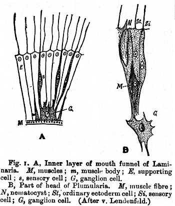
I. Comparative Anatomy and Phylogeny. In the Protozoa there is no differentiated nervous system, the fundamental nervous function -- IRRITABILITY (q.v.) -- being diffuse. In Sponges, though they are little more than colonies of protozoans, a few instances (Dendrilla, &c.) are known where definitive nerve-cells and sense-organs occur. In Coelenterata for the first time a well-developed nervous system appears, though the nervous function is not entirely concentrated within it. Especially interesting are the variations in the nervous system in different individuals of the colonies of Hydroids, consisting of nutritive individuals, blastostyles, and medusae. See Fig. 1.
The brains of lower vertebrates not only serve as simplified diagrams to explain the complex structure of the human brain, but afford hints as to the course pursued in its evolution. In Amphioxus we have at once the lowest existing type and a degenerate condition of the vertebrate nervous system. See Plate A (NERVOUS SYSTEM), Fig. 1. Here the cephalic portion of the nerve-tube is expanded, and indications of the three embryonic vesicles appear. The first of these forms a ventricle provided with an infundibulum connected with the olfactory apparatus. In the Petromyzontidae a marked advance upon the condition just described is reached. Here rudimentary hemispheres are developed, though they seem to be almost wholly concerned with data from the olfactory sense. The dorsal system of sense-organs (epiphysial system) is remarkably developed, suggesting a probable functional condition prior to the differentiation of lateral eyes. See PARIETAL ORGAN. The brain of selachians differs from that of higher vertebrates, in that no true lateral ventricles are formed. The lung fishes, although deviating less from the main line of evolution, are in some respects higher than sharks. The ganoids are still more specialized, forming a transition to the bony fishes. The cephalic end of the brain-tube connects with the ectoderm in an embryonic period, and the neuropore so formed is closely related with the rudiment of the hypophysis. In spite of great diversity otherwise, all bony fishes agree in lacking the cortex cerebri, and such nervous elements as substitute for that structure remain in the axial lobe. A dorsal membranous pallium covers the ventricles and aula, and includes the representatives of the plexus. The roof of the aula is distended to form a large dorsal sac. The infundibular region is also distended to form a saccus vasculosus near the mammillary bodies. Enormous massive protuberances from the pes pedunculi region constitute the hypoaria. The cerebellum is enormously developed, and a special portion, or volvula, is thrust into the unobliterated cavity of the mesencephalon. In addition to the cerebellum, other outgrowths from the medulla usurp positions in its roof, and these depend on the preponderance of one or another of the cranial nerve nuclei.
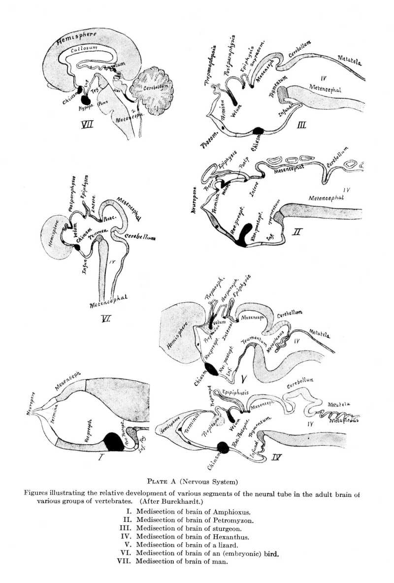
In all these groups a common structural plan appears in the midst of conflicting tendencies (cf. BRAIN). The figures from Burckhardt (Plate A, Nervous System) may serve instead of further verbal comparisons.
Literature: L. EDINGER, Vorlesungen ü. den Bau d. nervösen Centralorgane des Menschen u. d. Thiere (5th ed., 1896; trans. by Hall, Philadelphia, 1899); R. WIEDERSHEIM, Grundriss d. vergleichenden Anat. d. Wirbelthiere (3rd ed., Jena, 1893), 230-367; R. BURCKHARDT, Der Bauplan des Wirbelthiergehirns, Schwalbe's Morphol. Arb. (Jena), iv. No. 2; C. L. HERRICK, Nervous System, Wood's Ref. Handb. of the Med. Sci., ix. Suppl. (1893); C. GEGENBAUR, Vergleichende Anatomie d. Wirbelthiere, i (Leipzig, 1898); GAUPP'S revision of Echer's Anatomie des Frosches (Braunschweig, 1899); FLATEAU and JACOBSOHN, Handb. d. Anat. u. vergleichenden Anat. des Centralnervensystems d. Säugetiere, I. Makroskopischer Teil (Berlin, 1899); GOLGI, Sulla fina struttura del sistema nervoso, Riv. di Freniat. (1875 ff.), and Recherches sur l'histologie des centres nerveux, Arch. Ital. de Biol. (1882-91). For the current literature consult especially the Bericht über die Leistungen auf dem Gebiet der Anatomie des Centralnervensystems, published annually by EDINGER in Schmidt's Jahrbucher. This contains critical summaries of the more important papers in Comparative Neurology.
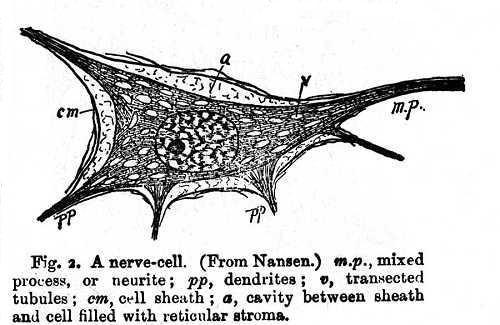
II. Histology and Histogenesis. The essential functional element of the nervous system is the nerve-cell (neurone or neurocyte) with its various appendages (see Fig. 2).
The nerve-fibre is simply a process or prolongation of the neurocyte or the product of the fusion of a number of such elements. A typical neurocyte consists of a cell-body, containing nucleus, nucleoli, pigmented and unpigmented protoplasm, and giving off at least two varieties of processes. One of these, which is usually the conveyer of cellifugal stimuli, is the neurite, 'axon,' or 'axis-cylinder process'; the others, usually more ramose, and conveyers of cellipetal stimuli, are the dendrites, or 'protoplasmic processes.' When a neurite soon breaks up without extending far from its origin, it is termed a neuropodium.
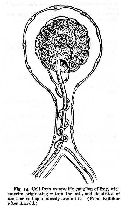
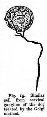
Some cells of the sympathetic ganglia form a peculiar type of unipolar neurocyte, in which the cell gives rise to a non-medullated fibre, and its body is surrounded by a dense network derived from a distinct fibre (Figs. 14 and 15). Heteropodal neurocytes are more generally distributed, forming the majority of functional nervous elements. Two main classes are distinguished: viz. type I, in which the neurite passes without interruption into a nerve-fibre, and type II, in which the neurite almost immediately subdivides in arborizations. Type I is best seen in the motor regions of the spinal cord. Type II is seen in sensory regions of the cortex, in the cerebellum, and in the retina.
Under most circumstances neurocytes are serially independent anatomically, i.e. do not actually come into contact with each other, thus insuring a certain insulation and rendering possible -- it may be conjectured -- a 'threshold' of discharge or an initial resistance. Cf. CONCATENATION (neural). In certain cases it would seem that there is lateral continuity among processes of cells of the same order (Dogiel). So in the retina and sensory elements of the skin, especially of the external genitalia. The perikaryon or cell-body and the neurite or fibre taken together make up the neural unit of structure called the NEURONE (q.v.), or neurocyte.
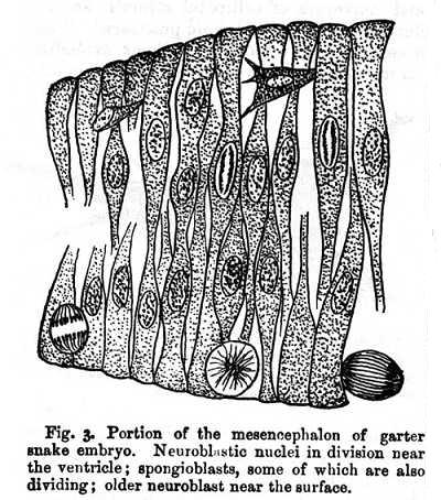
The same process occurs elsewhere in the brain, and it is probable that cells capable of proliferation are present as reserves up to a late period.
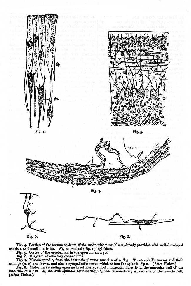
The simplest form of connection between peripheral and central elements found among vertebrates is illustrated by the olfactory connections (Fig. 6). The receptive cell (o) transmits its impulse along the neurite (n') to the olfactory glomerule (gl.) in the olfactory bulb. Here it passes over into the second nerve-unit, and thence via the neurite (n'') to the olfactory centres of the cortex. A more highly differentiated condition is found in the sensory nerves of the skin. See SPINAL CORD, Figs. 2 and 4. Here the cell-body of the first neurocyte has been removed from the surface to the spinal ganglion, and the central termini are much more complicated. These instances may be taken as typical intercellular relations.
III. Structure:
A. Central Nervous System. See BRAIN, and SPINAL CORD.
B. Peripheral Nervous System.
I. Dermal and Special Sense Organs. See SENSE ORGANS.
2. Nerves.
Nerve: a bundle of nerve-fibres, together with the sheaths and connectives. Each nerve-fibre is an outgrowth of some neurocyte or ganglion cell or the product of fusion of a moniliform series of such outgrowths. See CONCATENATION.
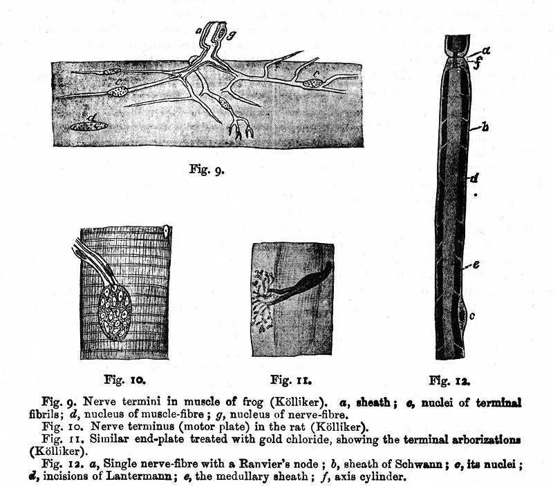
Each medullated nerve-fibre is divided into segments, each of which has its own nucleus (Ranvier's nodes). Phenomena of regeneration and development seem to indicate (contrary, however, to the statement of Kölliker) that each node represents a nervous element or cell, whose protoplasmic outgrowth (axis cylinder) has united with its neighbour in both directions. When the fibre is separated from its trophic centre, these nuclei proliferate and regenerate the fibre, supplying local trophic centres until the connection is re-established. The nodes also appear prior to or in the absence of the sheath of Schwann. A second obscure fragmentation of the myelin sheath gives rise to the Schmidt-Lantermann incisions, but these are probably artifacts.
Each nerve bundle is held together by an intrinsic connective system (endoneurium), and each bundle is covered by its epineurium. The sheath of the nerve as a whole constitutes its perineurium. See Fig. 12.
Motor nerve-fibres arise from neurocytes within the neuraxis and are thus prolongations of their neurites. Sensory fibres arise from the dendrites of the ganglion cells of the spinal or cranial ganglia, while the neurites of the latter enter the neuraxis, there to arborize about the cells of the terminal nuclei. The growth in each case pursues the direction of the normal nervous transmission. Cf. WALLER'S LAW.
Top the law that nerves conduct only in the direction of original growth there are exceptions, as in the case of the peripheral process of the spinal ganglion cells and as in cases where a sensory nerve has been grafted upon a motor trunk, and vice versa. The latter cases are probably to be explained on the supposition that the peripheral portion undergoes the characteristic DEGENERATION (q.v.), and, in regenerating, assumes the required properties.
Nerves are acted upon by mechanical, thermal, chemical, and electrical stimuli, though it is probable that each such irritant is converted into a common type of stimulus before it is translated through the nerve substance. The nature of the physiological or normal stimulus is still unknown (cf. END-ORGAN). The degree of excitability is largely dependent on the nutrition or the metabolic state. That which is frequently included under the term temperament (largely metabolic predisposition) probably has much influence.
The termini of motor nerve-fibres upon the muscle assume a variety of forms. The naked axis cylinder frequently branches extensively at its extremity, and even where definite end-plates are formed (Fig. 10), proper methods demonstrate similar ramifications among the nuclei (Fig. 11), which correspond to the terminal arborization of the motor nerve. See also Figs. 8, 9. Among the muscle-fibres a few of smaller size are found, usually in groups of several together, enclosed in a special connective-tissue sheath. The muscle-spindle, as such special muscle bundles are termed, is undoubtedly a sensory organ, and its fibres are supplied by nerve-fibres, which arise from the dorsal roots of the spinal nerves. Their peculiar spiral endings are shown in Fig. 7. The tendon is supplied with a special end-organ consisting of numerous fine branches. For the relations of the nerve roots see SPINAL CORD.
Literature: A. V. KÖLLIKER, Gewebelehre des Menschen, ii. Pt. I (1893); E. A. SCHÄFER, Histology of the Nerves, in Quain's Anatomy, i. Pt. II (10th ed., London and New York, 1895, with bibliography); L. GEDOELST, Étude sur la Constitution cellulaire de la Fibre nerveuse, La Cellule, iii (1887; contains a bibliography of 269 titles); also Nouvelles Recherches sur la Constitution de la Fibre nerveuse, La Cellule, v (1889); J. BEARD, The Development of the Peripheral Nervous System of Vertebrates, Quart. J. of Microsc. Sci., Oct., 1888; C. L. HERRICK, J. of Compar. Neurol., iii (March, 1893); AXEL KEY and RETZIUS, Studien in der Anatomie des Nervensystems und des Bindegewebes (Stockholm, 1875), 2 vols. folio; also Arch. f. mikr. Anat., ix (1873); L. RANVIER, Leçons sur l'Histol. du Syst. nerveux (1878); A. CHARPENTIER, Arch. de Physiol., [5] vi. 4. 792; G. CARL HUBER and LYDIA DE WITT, J. of Compar. Neurol., vii. Nos. 3 and 4 (1898), 169; also ibid., x. 160 (1900).
Cranial Nerves. The first twelve pairs of nerves leaving the central nervous system, and in higher vertebrates all emerging through special foramina in the skull. The last cranial nerves resemble the spinal nerves. This resemblance diminishes as we pass cephalad, yet there is little doubt that all cranial nerves have been differentiated from a similar primitive condition. There is, however, as yet no agreement as to the number of primitive segmental nerves represented in the head.
The current numerical designations, as given below (those of Sömmering, 1778), are highly unsatisfactory, though a better system has not yet been developed. The basis for a more philosophical classification is to be sought in the study of the components of the several nerves as determined by their central connections and peripheral distribution. All nerves arising from the same or homologous centres, and distributing to homologous peripheral organs, may be regarded as constituting a single system. In higher vertebrates, at least, there is no nerve which contains all of the components, and it is not necessary to assume that they were all contained in the primitive segmental nerve.
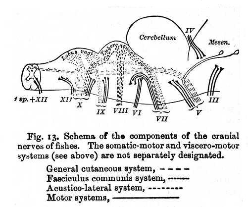
Gaskell has made an analysis of the nerves, in which he distinguishes five groups. His system is very suggestive, and affords a basis for further research. The reader is referred to his papers for the details (see the works cited below). Its most valuable feature is the analysis of the motor cranial nerve-nuclei into somatic (XII, VI, IV, III) and visceral (XI, X, IX, VII, V), a distinction based on structural, physiological, and embryological data (cf. Spinal Nerves, below). A more sure basis for the components has been laid, however, by their study in the lower vertebrates. They have been already worked out for some of the amphibians and fishes (O. S. Strong, 1895, et al.). The following systems of components are known in these forms, and there are probably others: (1) The general cutaneous system (corresponding to Gaskell's somatic sensory, in part). Sensory fibres carrying tactile and other general sensory impressions from the skin to the brain and terminating in the spinal trigeminal tract, which is the continuation into the medulla of the dorsal cornua of the spinal cord. The spinal trigeminus is not only a tract of fibres, but it contains cells scattered through it. These cells constitute the upper and lower sensory nuclei of the trigeminus. In the trunk this component is undoubtedly represented by most of the fibres of the dorsal roots of the spinal nerves. (2) The communis system. Sensory fibres carrying certain visceral and other special sensations (taste, &c.), often from more or less highly specialized organs (end-buds), and passing by way of the fasciculus communis of the medulla oblongata (fasc. solitarius of man) to the chief vagus nucleus or its equivalents (lobus facialis and lobus vagi of fishes). (3) The acustico-lateral system. Sensory fibres from the neuromasts, or sensory organs of the lateral line canals of fishes, and from the organs of the internal ear, which have probably been derived from the lateral line organs. In higher (amniote) vertebrates this system is represented only the auditory nerve. The fibres pass into the tuberculum acusticum of the medulla, a cluster of cells which, in lower forms at least, is intimately related to the cerebellum. It lies further dorsally than any of the other systems, and, unlike the general cutaneous, it seems to have no homologue in the spinal cord. Finally, the motor systems. Of these there are probably at least two, innervating (4) the somatic or body muscles, and (5) the visceral muscles respectively. They arise from the motor nuclei of the medulla. The fourth system is derived from the ventral cornua of the spinal cord or the cranial nuclei corresponding to them; the fifth probably from the lateral cornu or its equivalents in the medulla. The olfactory and optic nerves cannot as yet be placed in any of these groups. In man the same general relations of components undoubtedly prevail, though with great variation in the details, the chief difference being the absence of the acustico-lateral component in the VII and X nerves, correlated with the loss of the lateral line organs. The accompanying figure (13) illustrates their relations, so far as known, in fishes and amphibians. The XI and XII nerves in these forms are not completely differentiated.
I. Olfactory Nerve. The specific nerve of smell. It exhibits numerous important differences from the other cranial nerves, especially in that its fibres lack the medullary sheath, and are in direct protoplasmic continuity with their peripheral receptive organs, the sensory cells of the olfactory epithelium. A further difference from other sensory nerves is the apparent lack of a root ganglion. It has been suggested that this ganglion is represented by the olfactory cells of the nasal membrane, which have retained the primitive position in the skin in which they are found in some invertebrates. Centrally, the olfactory fibres break up into terminal arborizations in the glomerulae of the olfactory lobe, and here connect with other systems of fibres, which bring the glomerulae into relation with the cortical centres for smell in the hippocampal regions (see Fig. 6).
II. Optic Nerve. The nerve to the lateral functional eyes of vertebrates. (For parietal nerve to the median eye see PARIETAL ORGAN, and EPIPHYSIS.) The peculiar relations of the retina to the brain give to the optic nerve a very special significance. In fact, it is not a nerve at all, in the strict sense of the term. The protons of the eyes having developed on the cephalic plate of the embryo before the closure of the medullary tube, they, together with the corresponding ganglia, are included by the invagination of the cephalic plate, and come to lie in the walls of the primary forebrain. This is their permanent position in the ascidians and Amphioxus, but in craniate vertebrates end-organs and ganglia are evaginated to form the optic vesicle from which the retina ultimately develops. The embryonic optic nerve is at first a tubular prolongation of the first cerebral vesicle. The optic fibres grow into it at a later period. Throughout life, in certain amphibia, it retains its lumen and spongioblastic framework like the brain proper. The cells of the third, or ganglionic, layer of the retina represent, perhaps, the root ganglion. From these cells most of the optic fibres arise and grow towards the brain, though a smaller number seem to grow in the opposite direction from brain to retina, there ending in free arborizations in the deeper retinal layers. These latter fibres probably transmit impulses centrifugally to the retina. In animals with eyes so placed that the field of vision of one eye does not overlap that of the other eye, the optic nerves cross completely in the chiasma before entering the brain. In other cases, as in man, fibres from that portion of the retina whose impressions come from objects on the opposite side of the body do not, like the other fibres, cross in the chiasma. In the lower vertebrates the fibres of the optic nerve pursue a perfectly simple course through the chiasma and optic tracts to end in the superficial layers of the tectum opticum. Those fibres which end free in the retina arise from cells which lie in the tectum. Connections with the cerebrum are effected by means of the brachia, corpus geniculatum, and optic radiations. In mammals the general plan is the same, though complicated by connections of the optic tracts with the corpus geniculatum externum and the pulvinar, as well as with the tectum.
IV. Trochlear or Pathetic Nerve. The motor nerve to the superior oblique muscle of the eye. Its nucleus of origin lies in the floor of the aqueduct immediately behind that of the oculomotor. The fibres take a dorsal course and cross in the brain roof (the valvula) just cephalad of the cerebellum.
V. Trigeminal or Trifacial Nerve. The largest of the cranial nerves. It supplies the muscles of mastication and the general sense-organs of the face and teeth. It arises by two roots. The portio minor, or motor root, springs from two groups of cells, one lying dorsally of the exit of the nerve from the medulla oblongata. A small number of fibres arise in an elongated cluster of large cells arranged sparsely along the side of the aqueduct, and constitute the descending, or mesencephalic, root. The portio major is a complex structure, chiefly sensory. Most of the fibres go to the spinal trigeminal tract and terminate in cells lying in its path (including the 'chief sensory nucleus of the trigeminus'), and are, therefore, homologous with those of the dorsal roots of the spinal nerves. These supply the general cutaneous sense organs of the face (first component). Other fibres are said to enter the cerebellum. The fibres of the portio major before leaving the cranium center the Gasserian ganglion, of whose cells they are the neurites and from which they emerge in three branches, the ophthalmic, maxillary, and mandibular rami. The fibres of the portio minor pass by the Gasserian ganglion and enter the mandibular nerve. The ophthalmic nerve (exclusively sensory) sends small filaments to the third, fourth, and sixth nerves, to the ciliary ganglion, and from the latter to the ciliary nerves of the eye. The remaining fibres distribute to the dura mater, nose, eyelids, and skin of the forehead. The maxillary nerve also communiates with a small ganglion, the spheno-palatine, and supplies the integument of the side of the face, the upper teeth, and parts of the lining membranes of the nose and mouth. The mandibular nerve communicates with the submaxillary and the octic ganglia, and sends motor branches to the muscles of mastication, and sensory branches to the lower part of the face, to the tongue, mucous membrane of mouth, lower teeth, salivary glands, &c.
VI. Abducens Nerve. This purely motor nerve arises from a nucleus in the floor of the fourth ventricle, and passes ventrally to its superficial origin on the caudal edge of the pons. It innervates the external rectus muscle of the eyeball.
VII. Facial Nerve. A mixed nerve, arising in two portions. Most of the fibres are motor, and come from a nucleus lying ventrally and slightly caudally of that of the sixth nerve. The fibres curve around the latter, forming the genu of the facial, then directly ventrad in a compact bundle. After emergence from the brain they are joined by the much smaller portio intermedia from the fasciculus solitarius (or fasc. communis -- second component). These are sensory fibres and are provided with a ganglion (geniculate g.). They communicate by means of the chorda tympani with the submaxillary ganglion and the lingual nerve of the trigeminus, and supply taste-buds of the tip of the tongue. The larger (motor) component of the seventh nerve supplies the muscles of the expression of the face. The facial nerve is said by Edinger to receive some general cutaneous fibres from the first component (spinal V tract). In fishes and amphibians the seventh nerve receives also a large contingent from the acustico-lateral system.
The cortical connections of the third to seventh nerves are not thoroughly understood. There is, however, abundant evidence that the sensory components are all in more or less direct connection with the fillet (and hence with all the higher sensory centres), and the motor with the pyramids.
VIII. Auditory or Acoustic Nerve. Our knowledge of this nerve is in a most unsatisfactory state. The anatomical and functional relations of the several roots in man are still largely matters of controversy. In lower vertebrates the auditory nerves and the nerves of the lateral line terminate together in the tuberculum acusticum (see Fig. 13). In terrestrial amphibians (frog) the lateral line system disappears, and the auditory fibres end in two groups of cells lying dorsally of the superficial origin of the nerve, the tuberculum acusticum. The intimate relation of the auditory to the lateral line nerve is not confined to their central connections; but the membranous ear is probably a more highly specialized portion of the original lateral line canal system. The eighth nerve has two branches, the ventral, or vestibular, and the dorsal, or cochlear. The vestibular ramus supplies the macula acustica utriculi and the superior and external ampullary organs, and carries a ganglion (vestibular g.). The cochlear ramus supplies the macula sacculi, the posterior ampullary organ, and the cochlea. It, too, bears a ganglion (spiral ganglion). The two rami remain distinct to their terminal nuclei in the medulla, the vestibular ramus passing, for the most part, directly to a dorsal nucleus in the floor of the fourth ventricle, and the cochlear ramus into the ventral cochlear nucleus. Some fibres pass through this nucleus, externally and dorsally of the restiform body, to form the striae medullares in the floor of the fourth ventricle; others along the ventral wall of the medulla (trapezoid body) to the superior olives. The fibres of the cochlear ramus enter by devious courses into relations with the lateral lemniscus, and through the latter with the post-geminum, geniculatum mediale, and temporal lobe of the cerebral cortex. The vestibular ramus communicates directly with the cerebellum. In man the dorsal (cochlear) ramus is clearly the one chiefly concerned with audition. In the lower vertebrates there is no anatomical evidence that the utricular ramus differs in any way from the saccular. The vestibular fibres are regarded by many as serving the function of equilibration rather than audition.
IX. Glossopharyngeal Nerve. This nerve is intimately related to the tenth. It contains three elements: viz. (1) sensory fibres of taste, terminating among small cells in the dorsal part of the medulla, and distributing peripherally to the mucous membrane and circumvallate papillae of the back part of the tongue (second component); (2) general sensory fibres (first component); (3) finally, certain fibres arise from a motor nucleus -- the nucleus ambiguous -- lying on the ventral side of the sensory nucleus. These fibres innervate the stylopharyngeus muscle, and perhaps some of the constrictors of the pharynx (fifth component). Besides the lingual and motor branches above referred to, there are a tympanic branch to the middle ear, several pharyngeal branches, and anastomoses with the V, VII, X, and sympathetic nerves.
X. Vagus or Pneumogastric Nerve. The motor and sensory nuclei of the tenth nerve are scarcely distinguishable from the corresponding nuclei of the ninth. The size of the first (general cutaneous) component varies greatly in different animals. In fishes and amphibians there is also a large root entering the vagus from the tuberculum acusticum which supplies the organs of the lateral line of the trunk. In man the vagus communicates with the VII, IX, XI, XII, 1st and 2nd spinal, and the sympathetic. Its distribution is very wide, comprising branches to the dura mater, external ear, pharynx, larynx, heart, lungs, stomach, and other viscera. Throughout its entire extent the vagus is in frequent communication with the sympathetic, and it seems to share many of its functions with the latter. It controls, more or less directly, the more important automatic and vegetative functions of the body, such as circulation and digestion, and is thus of the most profound physiological significance.
XI. Spinal Accessory Nerve. The eleventh nerve consists of two parts: (1) The vagal portion is really detached filaments of the vagus, containing inhibitory fibres for the heart, motor fibres for the pharynx, &c. (2) The spinal portion arises from the lateral aspect of the spinal cord as far back as the fifth or sixth cervical vertebra in numerous separate strands. The fibres are motor, and arise from the lateral cornu of the cord. They turn cephalad, and are collected into a single trunk and emerge through the same foramen as the vagus. They supply the sterno-mastoid and trapezius muscles.
XII. The hypoglossal nerve arises from a large nucleus lying ventro-laterally of the central canal and chiefly below the fourth ventricle. The fibres are motor, and innervate the muscles of the tongue chiefly.
Literature: for further anatomical details consult the general works referred to under BRAIN, especially QUAIN'S Anatomy (10th ed.), iii. Pt. II, and Handatals der sensiblen und motorischen Gebiete der Hirn- und Rückenmarksnerven, by C. HASSE, Wiesbaden (1895). For a general critical summary of all that has been written on the comparative anatomy of the cranial nerves, and especially on the relation between cranial and spinal nerves, see MAX FÜRBRINGER, Ueber die spino-occipitalen Nerven d. Selachier u. Holocephalen u. ihre vergl. Morphol. (1897). On the components the following will be found useful: OLIVER S. STRONG, J. of Morphol., x. 1 (1895); B. F. KINGSBURY, J. of Compar. Neurol., vii. 1 (1897); C. JUDSON HERRICK, J. of Compar. Neurol., ix, and Arch. of Neurol. and Psychopathol., ii (1899); W. H. GASKELL, J. of Physiol., vii, x; VON KUPFFER, Merkel und Bonnet's Ergeb., 562-618 (1896). On sensory fibres of the eye-muscle nerves see C. S. SHERRINGTON, Proc. Roy. Soc. London, 1xi, No. 373, p. 247 (1897); G. C. HUBER, 'Atypical Motor Endings' of Retzius, Anat. Anz., xv (1899); and J. of Compar. Neurol., x. 2 (1900).
Spinal Nerves. There are in man thirty-one pairs of spinal nerves. Each pair, except the first, is named after the vertebra below which it emerges; thus we have eight cervical, twelve thoracic, five lumbar, five sacral, and a coccygeal nerve. Each spinal nerve arises by two roots from the SPINAL CORD (q.v.), of which the dorsal (sensory) is ganglionated. The ventral (motor) joins the ganglion, but merely to pass through it. Beyond the ganglion the mixed trunk divides into a small dorsal and a larger ventral ramus, each containing both sensory and motor fibres. At each segment there is a communication with the sympathetic ganglion of this segment.
It was formerly held that the spinal nerves contain but two components -- motor and sensory; but there is rapidly increasing evidence for separating the visceral nerves (efferent and afferent) from the somatic. We should therefore distinguish four kinds of fibres in each segmental nerve: (1) somatic afferent, sensory nerves from the skin; (2) somatic efferent, motor nerves for the voluntary, or skeletal muscles; (3) visceral afferent, nerves of 'general sensation,' &c.; (4) visceral efferent, supplying the involuntary muscles. The visceral systems are related to the lateral cornu and Clarke's column of the spinal cord. In the head these components are obscured, and, possibly, in part, supplanted by others; and it is not at present possible to establish sure homologies.
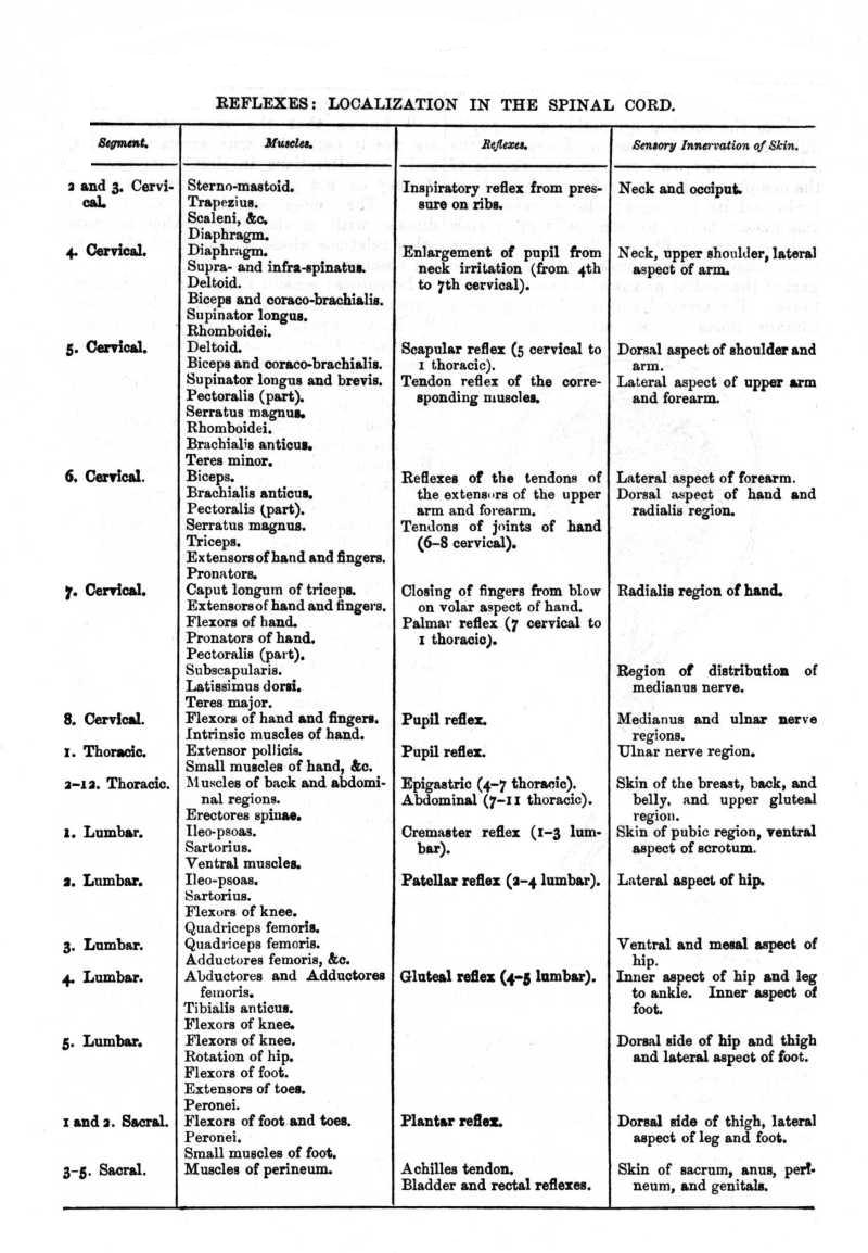
The table on the next page, from Edinger (based on Starr), exhibits the regional distribution from the various segments.
Literature: see the papers by GASKELL, cited under Cranial Nerves above, and QUAIN'S Anatomy (10th ed.), iii. Pt. II. 381-91 (with literature). For the courses of the several spinal nerves consult the textbooks of human anatomy already cited.
Sympathetic System. More properly, 'Sympathic System.' The sympathetic or visceral, as distinguished from the cerebrospinal nervous system, is primarily concerned with the vegetative functions. It consists of a double series of ganglia connected inter se and with the spinal ganglia, as well as with terminal ganglia adjacent to or imbedded in the visceral tissues.
The ganglia of the great sympathic chain (truncus sympathicus) are segmentally arranged, and each communicates with the corresponding spinal ganglion. The commissural fibres are of two kinds: (1) white (medullated), arising from the medulla oblongata by both dorsal and ventral roots, and terminating in the sympathic ganglia or passing through into the sympathic rami. They are both efferent and afferent. (2) Gray (non-medullated). They arise in the ganglion cells of the sympathic chain and join the spinal nerve just beyond the spinal ganglion. Some fibres turn peripherally into the spinal nerve; others terminate in the spinal ganglion around the bodies of the ganglion cells; others pass into the dorsal spinal roots and end in the sheath of that nerve, the dura mater, and the tissues adjacent, without entering the brain.
In the cervical and cephalic region the symmetry of this arrangement is disturbed, and the sympathetic fibres are bound up with the cranial nerves in the most complicated manner. In the abdominal cavity large ganglionic plexuses lie adjacent to the viscera. Of these the most important are: (1) the cardiac plexus, lying against the aorta and pulmonary artery. It receives fibres from the tenth cranial nerve. (2) The solar, or epigastric plexus, in the upper part of the abdomen, behind the stomach, is the largest. It communicates freely with the whole visceral innervation, and with several smaller secondary plexuses.
The vaso-motor nerves arise in the central system, but lose their sheaths in the sympathetic ganglia, and are thence distributed to the blood-vessels. See VASO-MOTOR NERVES.
Some sympathic functions are independent of central innervation; such are the automatic ganglia of the heart, and the plexuses of the intestine and uterus. Others are more or less indirectly or incompletely under central control. Among the important sympathic functions are, pupil reflexes, secretion, perspiration, digestion, and nutrition.
Conflicting views exist as to the origin of this system. Paterson claims that, so far from being derived from the central system, it is not even of ectodermal origin, but arises as an unsegmented mesoblastic cord. It is, however, more commonly regarded as an offshoot from the central system, and the evidence is in favour of the supposition that the sympathetic ganglia arise from neuroblasts that wander from the rudiments of the spinal ganglia and those of sensory ganglia on the cranial nerves. These cells are at first apolar and proliferate by karyokinesis. His and others show that there is an actual migration of such neuroblasts. The ganglionic centres associated with the visceral organs are in turn the results of similar migrations from the chain of sympathetic ganglia.
The fibres of the sympathetic are usually non-medullated. The cells, unlike those of the spinal ganglia, are usually multipolar; very different, however, are the peculiar spirally wound cells shown in Figs. 14 and 15. Small cells in the stroma of glands and in the plexuses of the intestines, or in the walls of blood-vessels, supply free dendritic termini to the muscular and epithelial structures in a wealth of detail until recently unsuspected.
Nervousness [Lat. nervosus, sinewy, vigorous]: Ger. Nervosität; Fr. nervosité, nervosisme; Ital. nervosismo (or neurosismo). A state of the nervous system characterized by an instability which may become variously manifest in conduct, emotion, and thought.
It is a frequent transitory experience in normal life, and is then often produced by a mixed group of predisposing causes, in which fear or apprehension is apt to be prominent. The apprehensiveness preceding an appearance before the public, the impatient awaiting of an announcement which may seriously affect one's welfare, the uneasiness or alarm provoked by a thunderstorm, the excitement of a patient before an impending operation may serve to suggest pertinent instances. The special sensitiveness to pain, and the anticipation of pain by imagination, are further nervous characteristics. Other symptoms of nervousness are motor restlessness, a tendency to start on slight alarm, palpitation, slight tremor, cold perspiration, tendency to be emotionally affected, excitability, irritability, and the like. Such moments or periods of nervousness may vary in intensity from slight deviations from normal equilibrium up to paroxysmal attacks of emotional disturbance, sometimes called 'nerve-storms.' The former may be considered as perfectly normal incidents under the influence of undue stress and strain, of critical periods of life, of ordinary fluctuations in physical and mental health. The latter are more or less pathological, and reflect an over-excitable condition of the nervous symptoms which, according to the accompanying systems and circumstances, may be recognized as neurasthenic, hysterical, epileptic, maniacal, &c.
Considered temperamentally, nervousness is a predisposition to states of instability of control; as such it plays a prominent part in the composition of human character as well as in the aetiology of mental disorders. Particularly in the problems of modern civilization, in the life that is lived under high pressure, with manifold demands on brain and sense-organs, on the emotions and the will, does the question of nervousness become of ever-increasing importance. The most direct pathological relation of nervousness is to NEURASTHENIA (q.v.); but it is also of fundamental importance in the aetiology of hysteria and other functional neuroses. The tendency to regard nervousness as equivalent to neurasthenia is not desirable, as the latter describes a typical disorder, while the former refers to a symptom or a temperamental characteristic which may accompany other disorders or may be quite normal.
Literature: see NEURASTHENIA; also KRAFFT-EBING, Nervosität u.
neurasthenische Zustände (1895); A. DE GIOVANNI, La Neurosi (1900). (J.J.)
Nescience [Lat. scientia, knowledge, + the negative prefix ne-]: Ger. Nichtwissen; Fr. nescience; Ital. nescienza. Literally, the condition of ignorance; but in a recent quasi-technical philosophical use, the theory that certain forms of reality (as God, the soul, matter in itself, &c.) are beyond our knowledge.
While often used as equivalent to AGNOSTICISM (q.v.), it is also employed to
describe the philosophy of Hamilton and Mansel, who would repudiate the title
of Agnostics, but who hold that only an indirect or mediate knowledge of the
existence of the Absolute, akin to faith or belief rather than to thought, is
possible. (J.D.)
Nestorians: Ger. Nestorianer; Fr. Nestoriens; Ital. Nestoriani. A sect of early Christians, founded by Nestorius, who maintained the individual distinctness of the divine and human elements in the nature of Jesus Christ, and consequently his bi-personality under the appearance of a unitary consciousness.
The doctrine of Nestorius roused a controversy which engaged the attention of a number of councils, and was finally settled by the banishment of Nestorius from Antioch, the Nestorian stronghold, about A.D. 433. The sect was transferred to Edessa, where it continued to flourish, spreading into Egypt, Arabia, India, and China. After the rise of Mohammedanism and during the Arabian domination in the East they increased their influence and stimulated an important intellectual development among the Arabs, the centre of which was at Bagdad. For several centuries they have been steadily declining, but still constitute several important communities of Eastern Christians.
Literature: SMITH and DWIGHT, Researches (1833); GRANT, Nestorians (1841);
DOUCIN, Hist. du Nestorianisme; ANDERSON, Hist. of Missions in the Oriental
Churches; WALCH, Gesch. d. Ketzereien; BAUER, Gesch. d. Dreieinigkeit; UEBERWEG,
Hist. of Philos., i, 'The Nestorians.' (A.T.O.)
Net [Lat. nitidus, clear, pure, through Fr.]: Ger. Netto; Fr. net; Ital. netto. Whatever part of a quantity remains after deducting the negative quantities which offset it. For instance, net income is an excess of income above expenses. 'If a person is engaged in business, his net income is found by deducting from his gross income the outgoings that belong to its production' (Marshall).
The conception of net income or earnings seems a simple one, but is really
very perplexing. What are the expenses involved in the production of a certain
article? Do they include the personal expenditures of the producer which fit
him for doing his work more thoroughly? Marshall, in his conception of public
net income or national dividend, does not include these things as business
expenses; but it is hard to give a reason for excluding them. On the other hand,
if we once begin to include any such expenses in our deduction, it is almost
impossible to tell where to draw the line. The best work in this subject has
been done by Cannan. (A.T.H.)