Distinguished Research Professor Doug Crawford sits down with Brainstorm to discuss the network of activity he and his PhD student discovered when researching how the brain perceives moving objects, how it achieves orientation and signals to the body to reach out and grab these objects. Understanding this network represents a major scientific advance.
Canada Research Chair in Visual-Motor Neuroscience, Distinguished Research Professor and Scientific Director, Vision: Science to Applications (VISTA), Doug Crawford is one of York University’s changemakers. For over 25 years, his work at the York Centre for Vision Research has focused on the control of visual gaze in 3D space, eye-hand coordination, and spatial memory during eye movements.
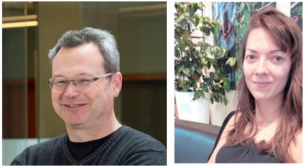
He’s also an academic who lets his students shine. With funding from the Natural Sciences and Engineering Research Council of Canada and the Canadian Institutes of Health Research, he recently produced an article in the Journal of Neuroscience with PhD candidate (biology) Bianca Baltaretu. She was a member of the Brain in Action International Research Training Group and did a research exchange with York’s German partners.

Crawford sits down with 'Brainstorm' to discuss the key findings of the 2020 article about reach-to-grasp movements and eye coordination.
Q: What were the study’s objectives?
A: The general question we’re interested in is: how does your brain keep track of everything that’s going on around you? You want to interact with the world when things are changing all the time in the external world. Also, your perspective changes as you move around.
Our key question is: how does the brain deal with it when you’re trying to grasp an object, and then the orientation of the object changes at the same time as you’re moving your eyes? This is a natural thing that happens in eye-hand coordination. But how does the brain keep track of this?
Q: This work builds on existing knowledge.
A: Yes. We know that some areas of the brain are involved in keeping track of the location of things when your eyes move. We know that some parts of the brain are involved in shaping of the grasp. We also, more recently, found some areas of the brain that keep track of the orientation of objects. We also know that an important part of the brain, called the parietal cortex, is involved. So that’s where we expected to see the action happening.
Q: Describe the experiment. You had 13 (human) participants and you measured their eye movements as they reached for a bar in complete darkness?

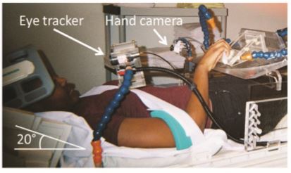
A: Yes, we measured eye position with an ‘eye tracker’ and hand position with a video camera when they were doing the task. We have an MRI machine to capture brain activity. There’s a sled-like apparatus from which participants can pull out of or put back into a magnet. The MRI allows you to measure blood flow in different parts of the brain. And we know that when the neurons are more active, it’s accompanied by increased blood flow. So, blood flow is an indirect measure of brain activity.
Participants reclined on the sled, as shown in the photograph. The head coil they wore helped to do the brain measurements once it’s inside the magnet. We had them start every trial with the hand at the same position on their abdomen. And then we presented them with an oriented object, rotated to the right or left. They fixated their eyes to the left or to the right when they saw this object. Then, in darkness, they had a couple seconds to plan their movement.
Half the time we saw participants move their eyes to the opposite side just before they reach; half the time we changed the orientation of the object. There were some different possibilities. And so, as I mentioned, we were really interested in what parts of the brain are compensating for those kinds of changes.
Q: What did you think would happen –was your hypothesis correct?
A: We had a strong hypothesis. I would say the most surprising thing was that we got exactly what we were looking for ... and that usually doesn’t happen.
We suspected that one part of the brain was keeping track of the change, changes in orientation across the eye movement. That was this area called SMG in the parietal cortex. Then there were other areas in our superior parietal cortex that are known to be involved in grasp. We found that all those areas were being modulated by the eye movements and the change in orientation.
We saw this network of connectivity where areas involved in eye movements, areas involved in grasp, keeping track of object orientation, they’re all talking to each other at the same time.
Q: This is a major scientific advancement.
A: Yes, the major scientific advance here is that we have these pieces of the puzzle. They form the next step in understanding the brain because, for many years, we’ve been studying this area or that area trying to understand what it does. But to really understand how the brain is working, we have to know how these different areas of the brain, and different neurons in those areas, are connecting to each other. They’re sending information to each other; they’re all working together. That’s the direction we’re going, I think: Getting away from very regional activity.

Q: What kinds of applications could this lead to?
A: This could inform complex problems of the brain, sometimes caused by brain damage. There are well-known problems, like spatial neglect, where the person completely ignores everything on the opposite side of the damage. There are conditions like optic ataxia, where people have trouble aiming or pointing.
What this more complex study could shed light on is something called constructional apraxia. That’s a deficit where people struggle when building things or doing spatial tasks where they have to manipulate objects and put things together. I think that could be a direction where we would go in terms of applying this to a patient population.
Q: York is a community of changemakers in neuroscience. Can you describe its evolution onto the world stage?
A: York has a long history of studying vision and visual behaviour. With our MRI and other technologies, we’ve really added to our understanding of how the brain contributes to visual behaviours over the last decade. It’s very important for translating that clinically.
At VISTA, we’re setting up new collaborations with rehabilitation clinicians and scientists. We want to translate this kind of knowledge into real-world applications. For example, we’re hoping to set up some collaborations with the Toronto Rehabilitation Institute [part of UHN]. We’re moving beyond diagnosis to potential rehabilitation strategies.
York is blossoming as a neuroscience centre, a major player in the Canadian and international stage, moving ahead with our ability to apply this knowledge.
To read the research article, go to the journal’s website. To learn more about Crawford, visit his Faculty Profile page. For more information on VISTA, consult the website. For more on the Centre for Vision Research, see the website.
To learn more about Research & Innovation at York, follow us at @YUResearch; watch our new animated video, which profiles current research strengths and areas of opportunity, such as Artificial Intelligence and Indigenous futurities; and see the snapshot infographic, a glimpse of the year’s successes.
By Megan Mueller, senior manager, Research Communications, Office of the Vice-President Research & Innovation, York University, muellerm@yorku.ca
