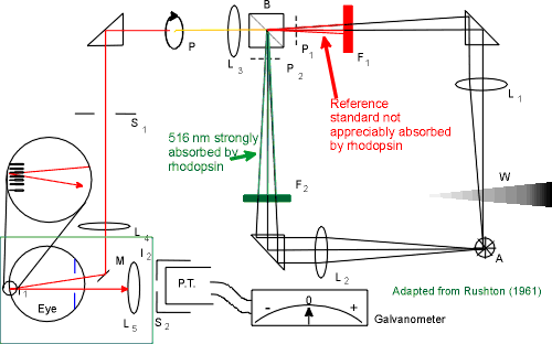
 This apparatus is used to measure the
amount of bleached rhodopsin in the outer segments of the rod
receptors. For a more complete explanation of this densitometer
the reader is referred to Rushton (1961).
At the start of the experiment the red and green filters (F1 & F2 ) were removed so that a very bright white light fell on the retina. This light bleached 100% of the rhodopsin which caused its appearance to change from a purple color to transparent.
Rhodopsin does not appreciably absorb deep red light so this color was used as a reference standard. Rhodopsin absorbs 516 nm very well so this was used as the test light. The two fixed polarizers (P1 and P2) polarized the red and green lights orthogonally. The rotating polarizer (P) caused the beam of light entering the eye to alternate between the reference and test fields. The neutral density wedge (W) controlled the amount of light in the reference field. The lights go into the eye and are reflected off the retina (see blowup), emerge from the eye and lens L5 projects the light into the photomultiplier tube (P.T.) and sends the signal to a galvanometer which provides a visual indication of the light reflected out of the eye.
As the rhodopsin regenerates it absorbs more of the 516 nm light thus reflecting less of it. The amount of the reference light is varied with neutral density wedge, W, so that the amount of light reaching the photomultiplier tube from the test and reference beams is equal. As the rhodopsin regenerates the more wedge has to be added to the reference beam to maintain a null reading on the galvanometer, thus indicating that the amount of each light is the same. By keeping track of the wedge position it is possible to plot values representing the amount of bleached rod photopigment.
Table of Contents
Subject Index
Table of Contents [When not using frames]