
The Larynx Part I: Overview and Cartilages
The Larynx serves a number of purposes. Though it may seem designed specifically for our speaking and singing, the larynx has evolved to allow us this control. It has other purposes too, ones that are essential to life. These purposes are called "biological", while speaking and singing are called "non-biological", as it is quite possible to survive without speech or singing. Witness those individuals who have had to have their larynges ( La - rin - jeeze,the plural of larynx) removed due to cancer and who talk through a process much like burping. Perhaps not very aesthetically pleasing, but possible.
Biological Function:
- to act as a valve to prevent air from escaping the lungs, e.g. weightlifting
- to prevent foreign substances from entering the lungs, trachea and glottis, e.g. while swallowing, the epiglottis covers the opening to the larynx.
- to forcefully expell foreign substances which threaten the trachea, e.g. coughing
Non-Biological Function:
- the production of sound
The images on this page can be viewed in a larger (slower to download) format by clicking on them.
Skeleton of the Larynx
gross features viewed from the front.
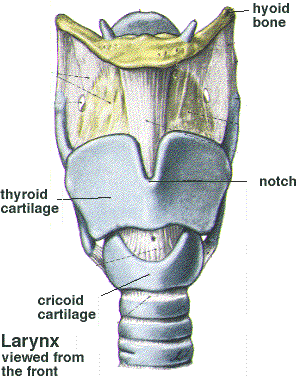
Hyoid Bone The yellowish bone in the image, it is horseshoe shaped and is the only bone in the body that floats, unconnected to another bone. It can be felt by pressing a finger into the crease where your chin becomes your neck.
Cartilages (visible in this image)
- Thyroid - the "adam's apple" on men, this V shaped cartilage features a notch in the front which can be felt with the edge of your thumb.
- Cricoid - a ring shaped cartilage connected to the trachea, it is larger in back where the arytenoid cartilages sit (not visible in this image).
Trachea
Made up of a series of cartilaginous rings, the trachea can stretch, much
like a vacuum cleaner hose. Compress it by swallowing, stretch it by tipping
your head back.
The Larynx, viewed from behind 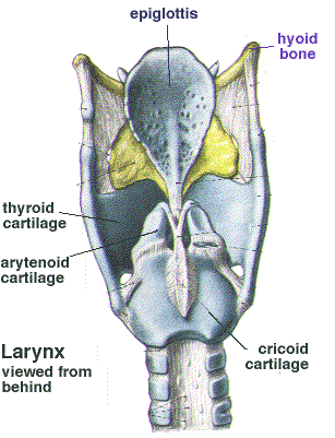
The Epiglottis
Functioning much like a "flap valve" on a toilet, the epiglottis
drops down in swallowing to close off the entrance to the larynx, thereby
protecting the airway.
The Fat Pad
Sitting behind the Epiglottis is a pad of fat (yellowish in the image above)
which cushions it as it rises.
The Arytenoid Cartilages
The arytenoids are pyramid shaped and sit on top of the widest part of the
cricoid cartilage. The vocal folds are attached to these cartilages and it
is their movement that opens and closes the glottis (the space between the
vocal folds).
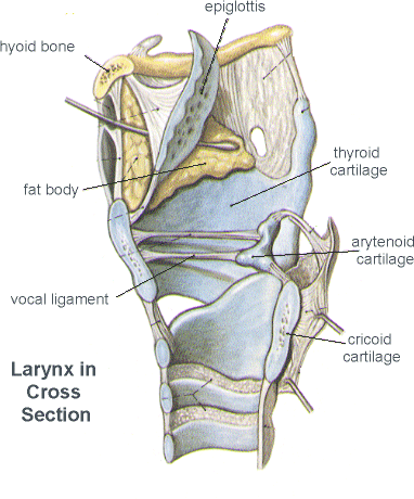 This image shows the larynx
from the side, featuring the vocal ligament, so that you can visualize the
placement of the vocal folds within the structure of the cartilages.
This image shows the larynx
from the side, featuring the vocal ligament, so that you can visualize the
placement of the vocal folds within the structure of the cartilages.
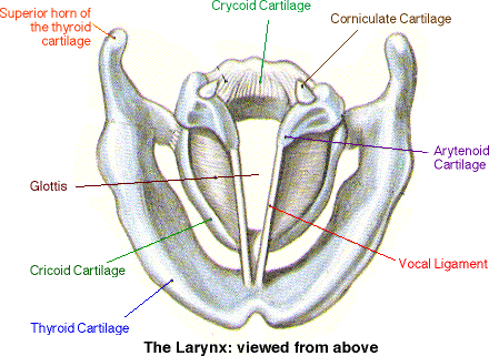 This image shows the cartilages
of the larynx from above, giving an excellent reference point for future images
of the larynx as seen through an endoscope, as they really appear.
This image shows the cartilages
of the larynx from above, giving an excellent reference point for future images
of the larynx as seen through an endoscope, as they really appear.
On to part two, Muscles and
Mucosa
Back to Phonation
More on Larynx
Anatomy
of the Larynx
by Dr Donal Shanahan, Anatomy
& Clinical Skills Centre, The School of Surgical Sciences at the University
of Newcastle-upon-Tyne. A fabulous use of web technology to teach the anatomy
of the larynx. Some of the best stuff I've seen. Highly recommended.
The
Voice-Centre at Eastern Virginia Medical School
A site dedicated to voice
and the larynx, this site has a few excellent pages on the larynx and its
anatomy. Highly recommended.
Anatomy
of the Larynx
The Gross Anatomy Course
at The University of Texas Medical School at Houston has a very in depth on
line resource from their 1997 labs. Designed for medical students, it is an
excellent source on detailed information beyond the scope of what is covered
here.
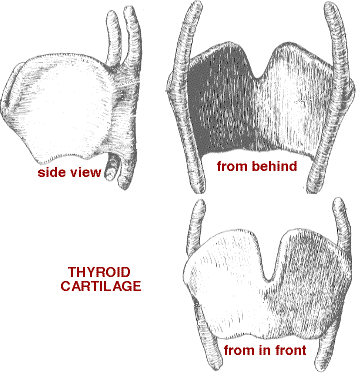
Thyroid Cartilage
An image of the thyroid
cartilage view from front, back and side.